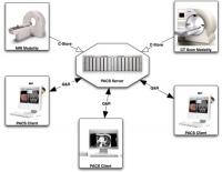
 Images from all modalities are accessible from any workstation
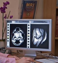
 CT images reveal hidden depths...
Modalities can include computed and digital radiography, MRI, CT and PET, ultrasound, gamma scintigraphy, and endoscopy

What is Dicom?
Dicom is a well-established IT standard used in many hospitals (human and veterinary) worldwide. Originally released in 1985 and regularly updated since, 3D ultrasound being one of the most recent additions, it is designed to ensure that systems from different manufacturers are interoperable and can exchange patient data easily.
Dicom defines the way medical images and patient data are produced, stored, displayed, processed, sent, queried, retrieved, and printed. Sadly, those responsible for defining the Dicom standards have never provided for ‘owner’ details to be recorded with patient information, humans generally not having owners to look after them! This gives rise to some incompatibilities between veterinary systems as organisations choose to handle this problem in different ways. Any such can usually be resolved quite easily within a PACS by re-mapping owner fields to match correctly.
How Dicom works in practice
Appointments booked for patients requiring diagnostic imaging are presented on the screen and the vet/radiographer/sonographer takes the images. These are automatically tagged with the patient information and added to the animal history. Workstations around the practice retrieve images from the PACS store, or can have selected images pre-fetched, for clinical diagnosis and/or review.
WORKLIST: when appointments are made and patient details noted, these can be presented to the vet or specialist who will carry out the procedure on the screen of the CR/DR, ultrasound, MRI etc computer. Vets do not have to type any details, just pick the correct patient from the scheduled list presented. As images are taken, these automatically have the patient data embedded in the file with the images, which the two can never be separated accidentally.
DICOM VIEWER: special viewing software is required to open a DICOM file. Many are available and each supplier of medical imaging equipment includes such software for at least one workstation when you purchase scanners or scopes. eFilm Lite is one of the most widely used applications for Windows PCs, often accompanying images produced on CD. For many users, OsiriX for Mac is one of the best DICOM viewers, handling the widest range of images/CDs.
PACS STORAGE: Picture Archiving and Communications Systems store images received from medical imaging equipment and provide a central facility for everyone in the practice. Images, complete with patient details, can be provided to any workstation via the network, on CD, via the internet or a secure VPN link between sites. Any image ever taken, any time, at the original quality. No more turning the office/ car upside down looking for last week’s x-rays or ultrasound scan!
Who benefits from Dicom?
Vets: better access to images and reports when DICOM standards are in practice, allowing them to make faster, more accurate diagnosis, potentially from anywhere on the internet.
Patients: obtain more effective treatment more quickly.
Clients: better outcomes for valuable/much-loved animals; shorter recovery times from less invasive diagnostic procedures.